The first photos proud expectant parents show off to family and friends—ultrasound images of a growing fetus in its earliest days—are underwhelming and indecipherable. Families long to see something more lifelike. And soon that may be possible.
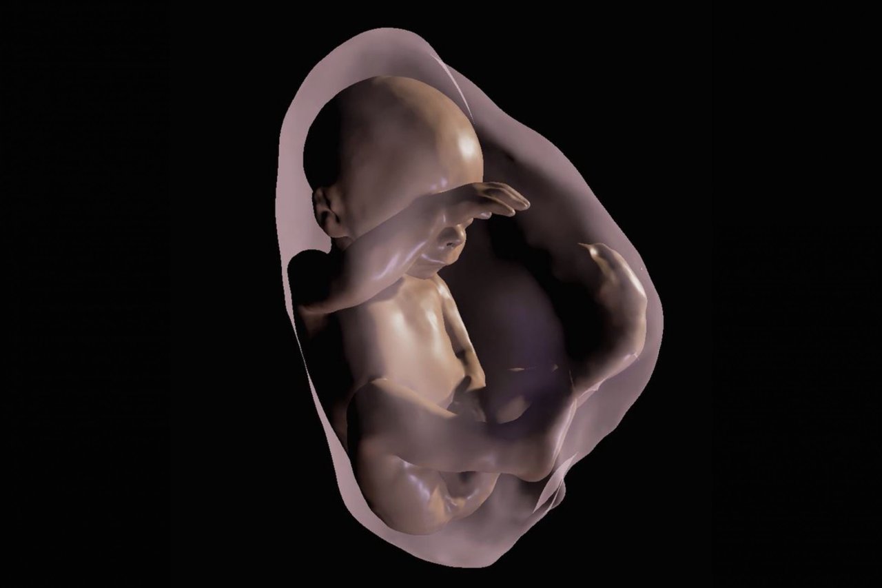
Researchers at Clinica de Diagnóstico por Imagem in Rio de Janeiro have developed a technology that can produce three-dimensional images of a fetus, umbilical cord and placenta using data from MRIs and ultrasound. With the aid of an Oculus Rift 2 virtual reality headset, parents will have a glimpse of their child in the womb before it enters the real world kicking and shrieking. The 3-D models are bound to start conversations about family resemblance before birth, but, more important, they could provide a way for physicians to diagnose structural abnormalities as early as the first trimester of life.
"We believe that these images will help facilitate a multidisciplinary discussion about some pathologies, in addition to bringing a new experience for parents when following the development of their unborn child," says Dr. Heron Werner, an expert in fetal medicine and lead researcher on the study.
With the headset, it's also possible to get a glimpse into the respiratory tract of the fetus. This allows the physician to see any life-threatening, abnormal mass blocking the airway. Eventually, the technology will provide 3-D views of vital organs, such as the heart. (Currently, the fetal heartbeat is audible through the headset.)
Werner, who presented his findings at the annual meeting of the Radiological Society of North America in November, has used the technology to produce models of 10 infants in utero, including ones with known congenital abnormalities. The goal, Werner says, is to develop a way to integrate the 3-D modeling technology into MRI and ultrasound machines so the 3-D likeness can be viewed much faster. Right now, producing the images takes some time, which isn't as useful in cases in which an infant might require emergency surgery or an early delivery.
Dr. Beth M. Kline-Fath, chair of the fetal imaging committee for the Society of Pediatric Radiology, says the models can help guide physicians monitoring pregnancies where the infant has anomalies of the face or mouth, such as large lesions, that make it difficult to breathe at birth. "Having a three-dimensional image could be helpful for planning at delivery to get a tube in the airway for support," she says.
And helpful, no doubt, for the newborn as well.