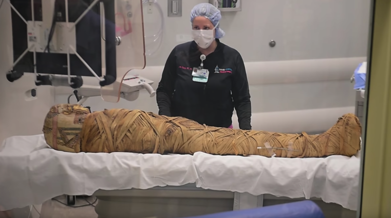
An imaging scan of a 2,000-year-old Egyptian mummy revealed a diagnosis doctors had been trying to pinpoint for more than a decade: cancer. Doctors recently subjected the mummified body of a man nicknamed "Hen" to a full computed tomography scan at the Crouse Hospital in Syracuse, New York, according to technology information site Boy Genius Report.
"He had a tumor on his fibula, which is one of the two bones of the lower leg," Dr. Mark Levinsohn of Crouse Hospital said in a syracuse.com video. "It had all the characteristics of a malignant tumor and one that's somewhat rare. So, here we have a rare circumstance and a rare tumor and that evoked our interest a lot."
This isn't the first time a mummy has been sent through a CT scanner; scientists at Northwestern University scanned the mummified remains of a young Egyptian girl just last month. And this actually isn't even the first time this mummy was sent through a CT scanner. Levinsohn said in the syracuse.com video that he first scanned Hen in 2006 in an attempt to make a diagnosis, but without success. Now, technology has advanced to the point where, as Levinsohn said in the video, they can bring their work to completion.
"Since that time, the last 10 years, they've upgraded the equipment," Levinsohn continued in the syracuse.com video. "What at that time was a 16-detector scanner is now a 320-detector scanner and all that additional information is now derived when we scan the body. So, we can tell all kinds of greater detail."
Those numbers refer to how many rows of radiation detectors—the core hardware that allows CT scans to image the inside of the body—the machine used. The more rows, the more detailed and accurate the image. The first CT scanners in the 1980s had only one row of detector arrays. The first machine with two rows was introduced in 1992. CT systems had 4 detector rows by the late 1990s, and now many machines, like the one at Crouse Hospital, have 320.
The scanner Levinsohn and his team used in 2006 was top-of-the-line at the time, he said in the video. But the 16-detector scanner didn't provide the level of detail they needed to make a diagnosis. A decade later, Levinsohn was finally able to see what he couldn't in 2006. Hen normally resides in the Cazenovia library in Syracuse, where library benefactor Robert Hubbard placed him in 1894 after purchasing him in Egypt, according to syracuse.com.
The modern CT scanners may be good, but we still don't know for certain whether Hen's ultimate cause of death was actually the cancer. As BGR wrote, any number of other fates could have befallen him before the disease progressed far enough to kill him.
"Some things will come out right away, but I think we won't be done with this until at least two or three more months," Levinsohn said in the video. He also said he considers Hen a community treasure.
Crouse Hospital provided the CT and diagnostic services pro bono, which is fortunate since Hen probably lacks a good insurance plan and is also extremely dead.
Uncommon Knowledge
Newsweek is committed to challenging conventional wisdom and finding connections in the search for common ground.
Newsweek is committed to challenging conventional wisdom and finding connections in the search for common ground.
About the writer
Kastalia Medrano is a Manhattan-based journalist whose writing has appeared at outlets like Pacific Standard, VICE, National Geographic, the Paris Review Daily, ... Read more
To read how Newsweek uses AI as a newsroom tool, Click here.