Most people have looked into a microscope in science class and seen cells crammed onto a glass slide, but new video footage shows cells and their movement in stunning detail to create a clear picture of how life works.
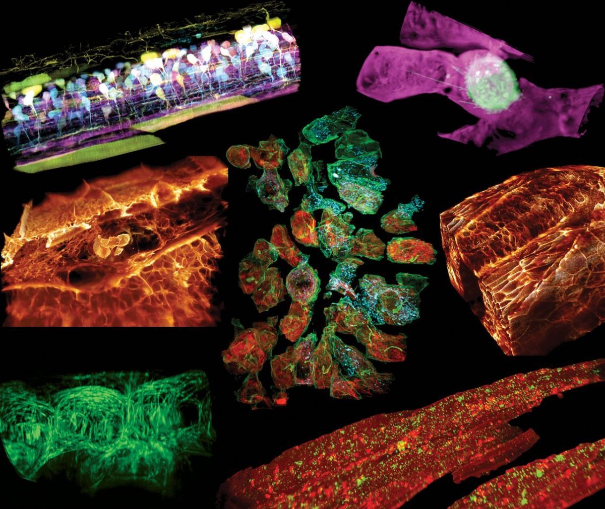
Researchers took images of immune cells from the ear of a zebrafish, which flit about like sparks of light, their flame-like shapes consuming small blue orbs that represent sugar particles. A second clip shows a green cancer cell oozing through a blood vessel, its extensions reaching out toward the vessel wall like a hairy trespasser. In another, a membrane in the eye of the fish pulses, its parts expanding and contracting. All of the video footage shows the microscopic pieces at work in real-time.
The video footage relies on a combination of two imaging techniques: adaptive optics, a technique astronomers use that employs reshapable mirrors to compensate for distortion in an image and gather more detail; and lattice light-sheet microscopy, which sends light through a field to create a bunch of 2D images, which are then woven together to construct a 3D one.
The researchers published their findings in the journal Science.
"It's often said that seeing is believing, but when it comes to cell biology, I think the more appropriate question is, 'When can we believe what we see?'" physicist Eric Betzig, one of the researchers, said in a statement from the Howard Hughes Medical Institute.
Without the advanced imaging techniques his team used to capture the cellular footage, that microscopic world is "just too damn fuzzy," Betzig said.
The old way of looking at cells under a microscope also loses the crucial component of seeing them act naturally, as they would interact with the rest of the body.
"Organisms live by means of the complex, dynamic, three-dimensional interplay between millions of components, from the molecular to the multicellular," according to the study. "Visualizing this complexity in its native form requires imaging at high resolution in space and time anywhere within the organism itself, because only there are all the environmental factors that regulate its physiology present."
Uncommon Knowledge
Newsweek is committed to challenging conventional wisdom and finding connections in the search for common ground.
Newsweek is committed to challenging conventional wisdom and finding connections in the search for common ground.
About the writer
To read how Newsweek uses AI as a newsroom tool, Click here.