Virtual reality can now literally get inside your head.
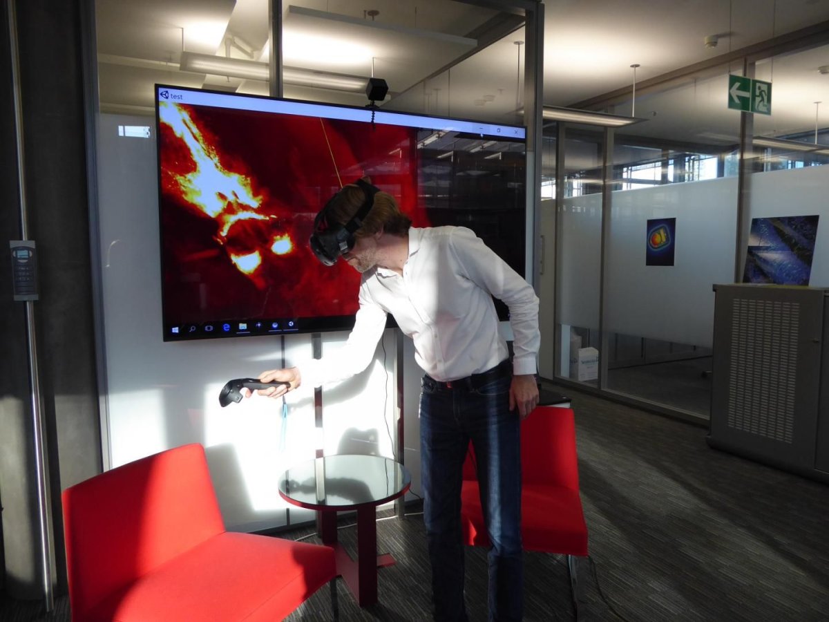
Researchers from the Wyss Center for Bio and Neuroengineering and the University of Geneva unveiled a virtual reality (VR) system designed to streamline analysis of brain-imaging data. The system will help doctors more efficiently wade through the sheer volume of images—and also sounds extremely cool.
By strapping on VR goggles, users can take a virtual-reality tour of the inside of a mouse brain. They can even interact with the data by highlighting or zooming in on the area of their choice using hand-held pointers. A poster outlining the system was presented today at the annual meeting of the Society for Neuroscience 2017, in Washington, DC.
"This new visualization technique could also be used to look inside a human brain," lead author Stéphane Pages, the staff scientist at the Wyss Center who managed the microscope fabrication, told Newsweek via email. "It's a quick and efficient way of analyzing large volumes of data from MRI scans."
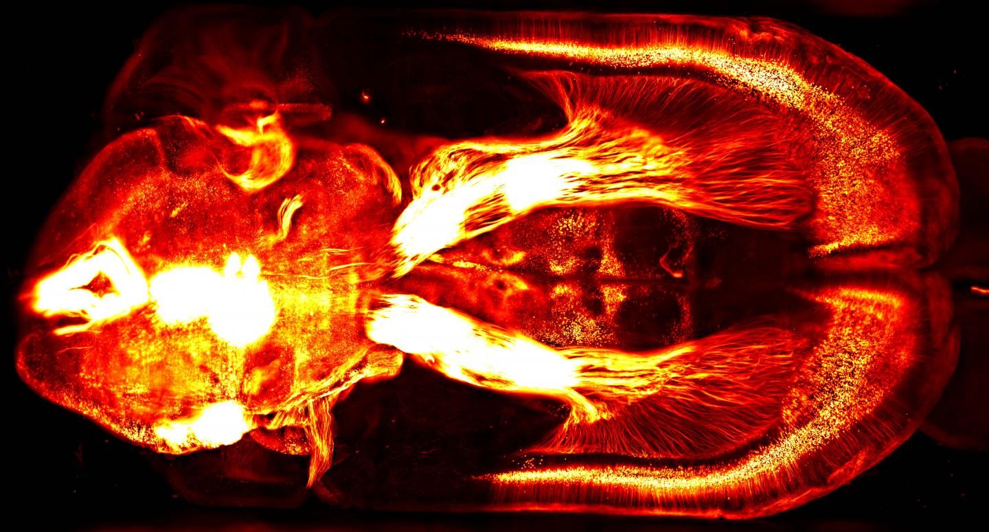
When the researchers injected a fluorescent compound into a mouse brain to trace activity in different areas, they were able to see the reactions involving dopamine; the circuits illuminated in the video are the ones linked to our brain's natural reward system. The new system also perfectly complements other cutting-edge tools like the Wyss Center's custom lightsheet microscope at Campus Biotech in Geneva, Switzerland.
A neuron is about five times thinner than a single human hair. The Wyss Center's lightsheet microscope is one of only three in the world sophisticated enough to image neurons individually. The researchers' recent work has already allowed the microscope to pick up enhanced details within the brain's circuitry.
The Wyss Center researchers expect that using the two systems together will have particularly exciting implications for their translational neurotechnology program. It will improve their ability to analyze the interactions between a patient's brain and any new medical implants that are attached to it. Plus, they can struggle less with the kinds of neural structures that are tricky for the human eye to pick up when they're flattened onto a screen; the VR system can image them instead.
"In the future, this could prove a very useful tool to gain a new 3D perspective on the complex mechanical and biological interactions between the human brain and new types of MRI compatible brain probes," co-author Gilles Reymond told Newsweek via email. "[Which] can be used to help people overcome nervous system disorders."
Uncommon Knowledge
Newsweek is committed to challenging conventional wisdom and finding connections in the search for common ground.
Newsweek is committed to challenging conventional wisdom and finding connections in the search for common ground.
About the writer
Kastalia Medrano is a Manhattan-based journalist whose writing has appeared at outlets like Pacific Standard, VICE, National Geographic, the Paris Review Daily, ... Read more
To read how Newsweek uses AI as a newsroom tool, Click here.