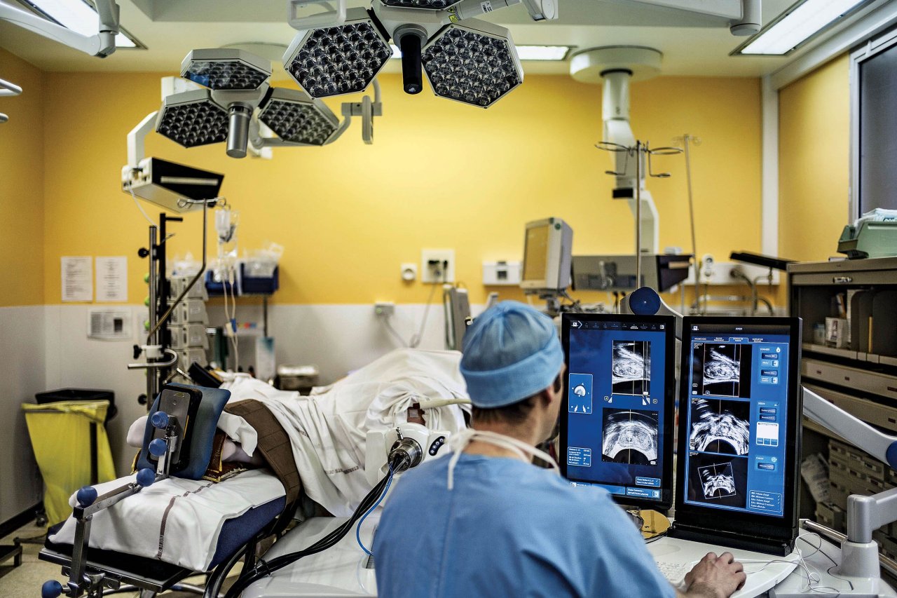
Medicine has come a long way from its "let's-just-open-it-up-and-take-a-look" past. Today, radiology provides doctors with a look into the body without the need for a single incision. Technologies like computer tomography (CT) and magnetic resonance imaging (MRI) have opened windows into vital organs, musculature and even the brain. But CT and MRI scans have limitations—they're 2-D images of body parts uploaded onto a computer, very different from real-life, 3-D organs.
"Doctors are trying to extract 3-D information from flat slices," says Sergio Aguirre, the founder and chief technology officer of EchoPixel, a tech company trying to solve the flat-screen problem. "We found they are looking at flat image slices, and correlating between images, and in the process, forgetting what they are looking at." He says that in their research, they found that doctors are trained to move from one 2-D image to the next, memorizing the orientation of each image as they go. But, he says, "they may forget a key feature that will lead them to lose important diagnostic information. It is a small mental lapse that's happening from data overload."
EchoPixel has designed medical visualization software that takes those 2-D slices, reconfigures the anatomy into its proper 3-D spatial structure and beams a depiction of the tissue and organs as if they were real physical objects hovering over a display board. The idea is to enable doctors (wearing 3-D glasses) to lift a skull off the screen with a hand-directed stylus, zoom in on aneurysms and freely rotate the hovering image so they can look at it from many angles. The software has potential in medical school, patient communication and, most important, diagnostic accuracy.
In training trials, radiologists using the software were able to cut their diagnosis time in visual colonoscopies from 30 minutes down to five to 10 minutes, and increased the detection rate of flat lesions—difficult-to-detect and often-overlooked precancerous tissue—by 20 percent. In another trial, Stanford University radiologists were able to use the 3-D images to generate improved surgical plans for major aortopulmonary collateral arteries, a heart condition, decreasing surgery time from four to 1.5 hours.
The 3-D platform was recently approved by the FDA for clinical use, and units have already been installed at Stanford University the University of California, San Francisco; Foxconn Technology Group; and Cleveland Clinic. The company says that the cost of the technology is comparable to—and in many cases, even cheaper than—the 2-D workstations currently considered the standard in health care facilities.
"There's no reason that, within a few years, anyone should be looking at a 2-D model," says Aguirre. EchoPixel is also about to begin a colonoscopy trial with real patients, and are working on a data-sharing system aimed at helping doctors across disciplines work together seamlessly on the same 3-D image sets. Universal adoption could have an untold impact on health care.
"All you have to do is look at how CT and MRI affected health care," says Dr. David Langer, chief of neurosurgery at Lenox Hill Hospital. "All of a sudden we saw things we never saw before, and we started to develop new strategies for treating problems we never knew about. [3-D imaging] could result in absolutely the same thing."